Abbreviations
This is a list of abbreviations used in the guide, sources from Elsevier https://www.elsevier.com/__data/promis_misc/JVCabbreviations.pdf and other sources in common use in veterinary echocardiology.
- A peak velocity of late diastolic transmitral flow (m/s)
- ACVIM American College of Veterinary Internal Medicine
- A-lines horizontal echogenic lines representing reverberation artefact seen when scanning lungs
- ANS autonomic nervous system
- Ao aorta
- AoD aortic root diameter
- Ao-Lax aortic root measured in long axis from the RPLx5ch
- AT acceleration time
- ATE aortic thromboembolism
- AV aortic valve
- B-lines comet-tail artifacts seen on lung ultrasound
- BSA body surface area
- BW body weight
- CFD colour flow Doppler
- CHF congestive heart failure
- CI confidence intervals, usually means 95%
- CKCS Cavalier King Charles Spaniel
- CVC caudal vena cava
- CW continuous wave Doppler
- DCM dilated cardiomyopathy
- DDx differential diagnoses
- DLVOTO dynamic left ventricular outflow tract obstruction
- E peak velocity of early diastolic transmitral flow (m/s)
- E:A ratio of E wave to A wave
- E:E’ ratio of E wave to tissue Doppler E wave
- E:IVRT ratio of E wave to IVRT measurement
- EF ejection fraction (%)
- EPSS E-point-to-septal-separation (mm)
- FAC fractional area change
- FS fractional shortening (%)
- HCM hypertrophic cardiomyopathy
- HR heart rate
- IVRT Isovolumic relation time (ms)
- IVS interventricular septum
- IVSd interventricular septum thickness at end-diastole (mm)
- IVSs interventricular septum thickness at end-systole (mm)
- LA left atrium
- LA:Ao left atrium to aortic root ratio
- LAA left auricular appendage
- LAD/LADmax maximal LA dimension from right parasternal long axis view (RPLx4ch)
- LAD:Ao maximal LA dimension from right parasternal long axis view ratio to aortic root
- LADn left atrial dimension normalised for body weight
- LAP left atrial pressure
- LAV/LAVn left atrial volume/left atrial volume indexed to body weight
- LV left ventricle
- LVEDV/LVESV LV end-diastolic volume/LV end-systolic volume (ml), often indexed to body weight as LVEDVn and LVESDVn (ml/kg)
- LVIDd/s left ventricular internal diameter in diastole/systole
- LVIDdN left ventricular internal diameter in diastole normalised for body weight
- LVIDsN left ventricular internal diameter in systole normalised for body weight
- LVET left ventricular ejection time
- LVFP left ventricle filling pressure
- LVOT left ventricular outflow tract
- LVFW left ventricular free (posterior) wall
- LVVd/s left ventricular volume at end-diastole/systole
- M mode motion mode, tissue motion collected from a single line graphed against time
- MAPSE mitral valve annular plane systolic excursion
- MV/MR/MD mitral valve/mitral valve regurgitation/mitral dysplasia
- MVD myxomatous valve disease, also known as myxomatous mitral valve disease, degenerative valve disease
- MPA main pulmonary artery
- MV/MR mitral valve/ mitral regurgitation
- PA pulmonary artery
- PDA patent ductus arteriosus
- PEP pre-ejection period
- PG pressure gradient
- PH pulmonary hypertension
- PI/PV pulmonic insufficiency/pulmonary valve
- PW pulsed wave Doppler
- RA/RV right atrium/right ventricle
- RAA/RAAn right atrial area/right atrial area normalised for body weight
- RAD right atrial diameter
- RPAD right pulmonary artery distensibility
- RR/RRR/SRR respiratory rate/resting respirator rate/sleeping respiratory rate
- RVDd/s right ventricular end diastolic diameter at end-diastole/systole
- RVFW right ventricle free wall
- RVOT right ventricular outflow tract
- S’ systolic motion in tissue Doppler imaging
- SAM systolic anterior motion (applied to mitral valve)
- SMOD Simpson’s method of discs
- TAPSE tricuspid valve annular plane systolic excursion
- TDI tissue Doppler imaging
- TMT transient myocardial thickening
- TV/TR/TD tricuspid valve/tricuspid regurgitation/tricuspid dysplasia
- VHS vertebral heart score (radiographic)
- VLAS vertebral left atrial size (radiographic)
- VSD ventricular septal defect
- VTI velocity time interval, a measure of mean velocity through the flow calculated by ultrasound machine from tracing the Doppler envelope
Main echo views
- RPLx4ch = right parasternal long axis 4 chamber view
- RPLx5ch = right parasternal long axis 5 chamber view, also known as left ventricle outflow tract view
- RPSx = right parasternal short axis views, at level of papillary muscles (‘mushroom’), mitral valve (‘fish mouth’), aortic root (‘whale and Mercedes Benz’) and heart base with pulmonic outflow
- LPLx4ch = left parasternal long axis 4 chamber view, also known as left apical view
- LPLx5ch = left parasternal long axis with LVOT
- LPCr = left parasternal cranial views
- SC = subcostal view, used to align with LVOT and aorta
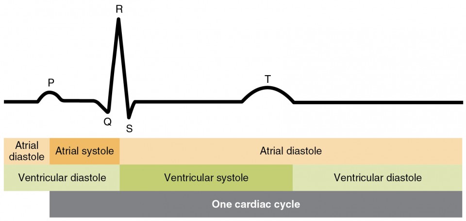
ECG